AI in medicine: can a selfie tell how long we might live?https://en.majalla.com/node/334184/science-technology/ai-medicine-can-selfie-tell-how-long-we-might-live
AI in medicine: can a selfie tell how long we might live?
Artificial intelligence can analyse facial features for clues to biological ageing and cancer survival, but further validation is needed before clinical use
Ibrahim Arafath
AI in medicine: can a selfie tell how long we might live?
In 2016, while visiting New York City, Megan Troutwine took a selfie and noticed one eyelid seemed unusually droopy. After returning home, she immediately consulted a neurologist. A subsequent magnetic resonance imaging (MRI) scan revealed a meningioma, a tumour that develops in the membranes surrounding the brain and spinal cord.
The photograph was not a diagnostic tool, and doctors did not use artificial intelligence to detect the tumour. It simply alerted Troutwine to a physical change that warranted medical investigation. Her experience illustrates how a chance photograph can draw attention to an unsuspected medical condition.
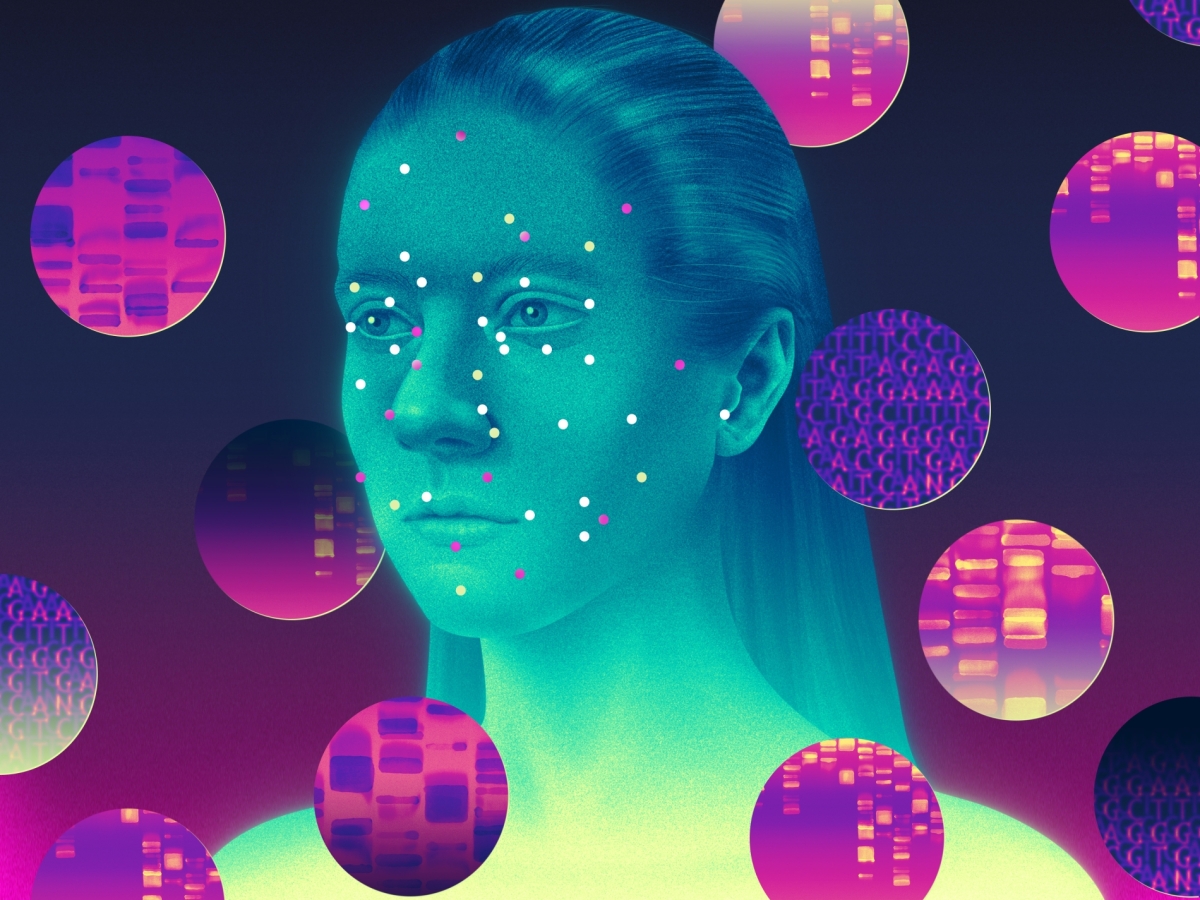
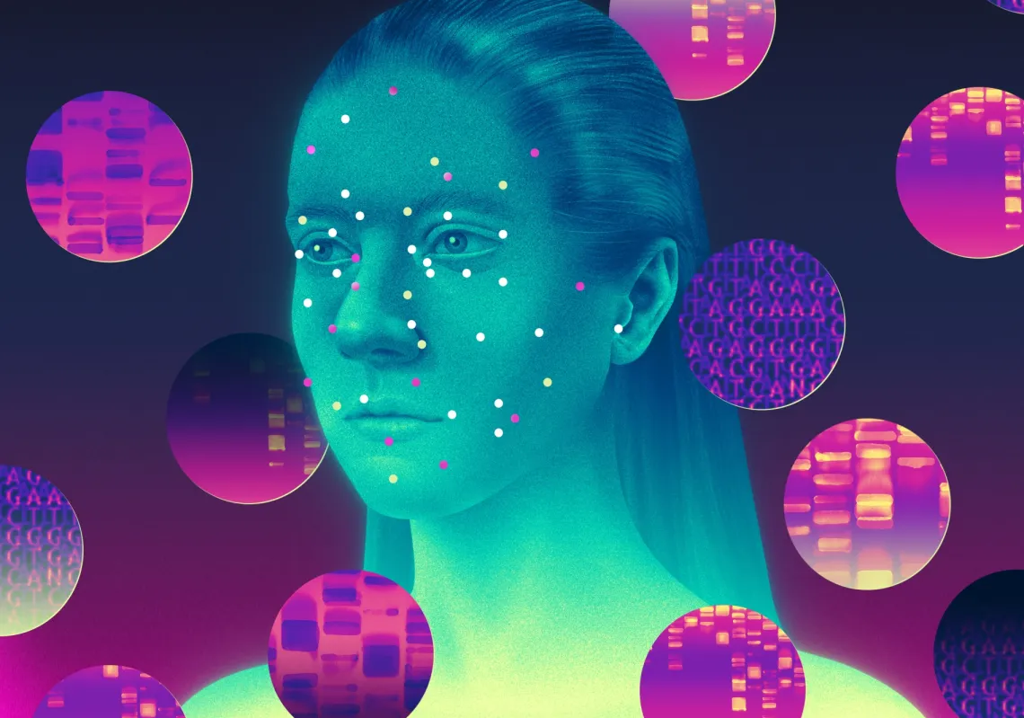
Subtle changes in facial features can sometimes reveal symptoms that warrant medical attention. Scientists are now looking beyond what the human eye can discern, developing artificial intelligence systems that analyse facial photographs for signs linked to health, biological age, and even survival prospects among cancer patients.
These advances suggest that personal photographs, now part of everyday life for billions of people, could become an additional source of medical information without the need for specialised imaging equipment or complicated procedures.
Yet identifying statistical patterns in facial features is a long way from establishing whether photographs can reliably diagnose disease or guide treatment. That's why researchers are now pursuing a more ambitious goal: using algorithms to identify visual patterns that may contain health information beyond the reach of ordinary observation.
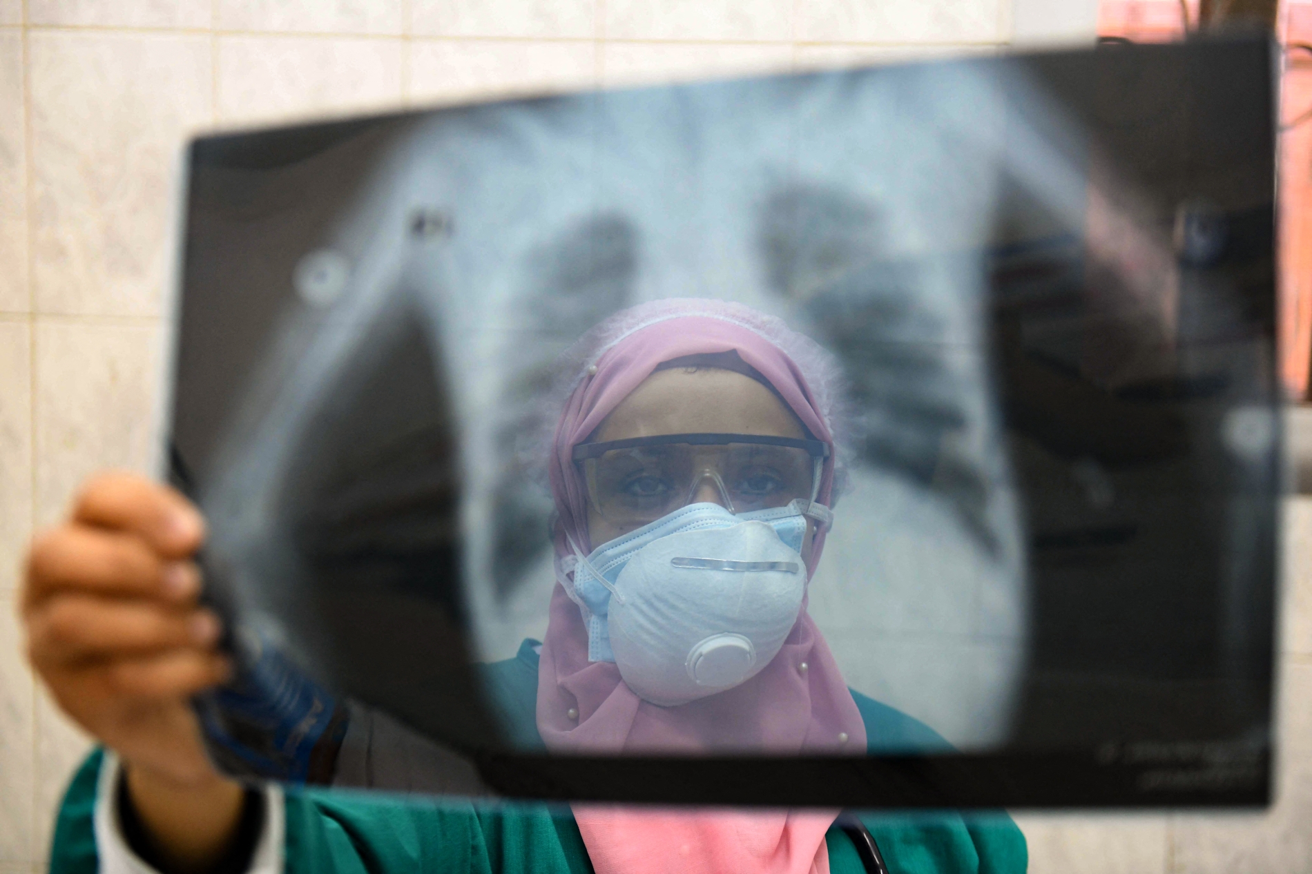
An Egyptian doctor wearing two protective masks checks a patient's lung X-ray at the infectious diseases unit of the Imbaba Hospital in Cairo.
Medical function
Photography has served medicine for years. As early as the 19th century, doctors used photographs to document physical abnormalities and monitor bodily changes associated with disease.
Today, smartphones equipped with high-resolution cameras and advances in digital communications have given photography a new role in healthcare. This role became particularly apparent during the Covid-19 pandemic, when hospitals and clinics reduced face-to-face appointments and increasingly relied on remote consultations.
The Countess of Chester Hospital in the UK, for example, used photographs patients took on their mobile phones to help assess skin lesions suspected of being cancerous and determine the appropriate course of care.
Identifying statistical patterns in facial features is a long way from establishing whether photographs can reliably diagnose disease or guide treatment
A study published in January in Annals of Plastic Surgery examined the hospital's experience between June 2020 and June 2021. It involved 939 patients, whose average age was 70.8 years. Doctors conducted telephone consultations supported by patient-submitted photographs. They assessed images for clarity, magnification, and the accurate identification of the lesion's location. The findings showed that 90.8% of patients were listed for surgery following the remote assessment.
That figure does not represent the diagnostic accuracy of photographs in detecting skin cancer, nor does it mean that all the patients were subsequently diagnosed with the disease. The images remained a supplementary tool, alongside medical examinations and, where necessary, the histological analysis required to confirm a diagnosis. However, researchers are now developing more sophisticated systems to extract and interpret medical information from such images.
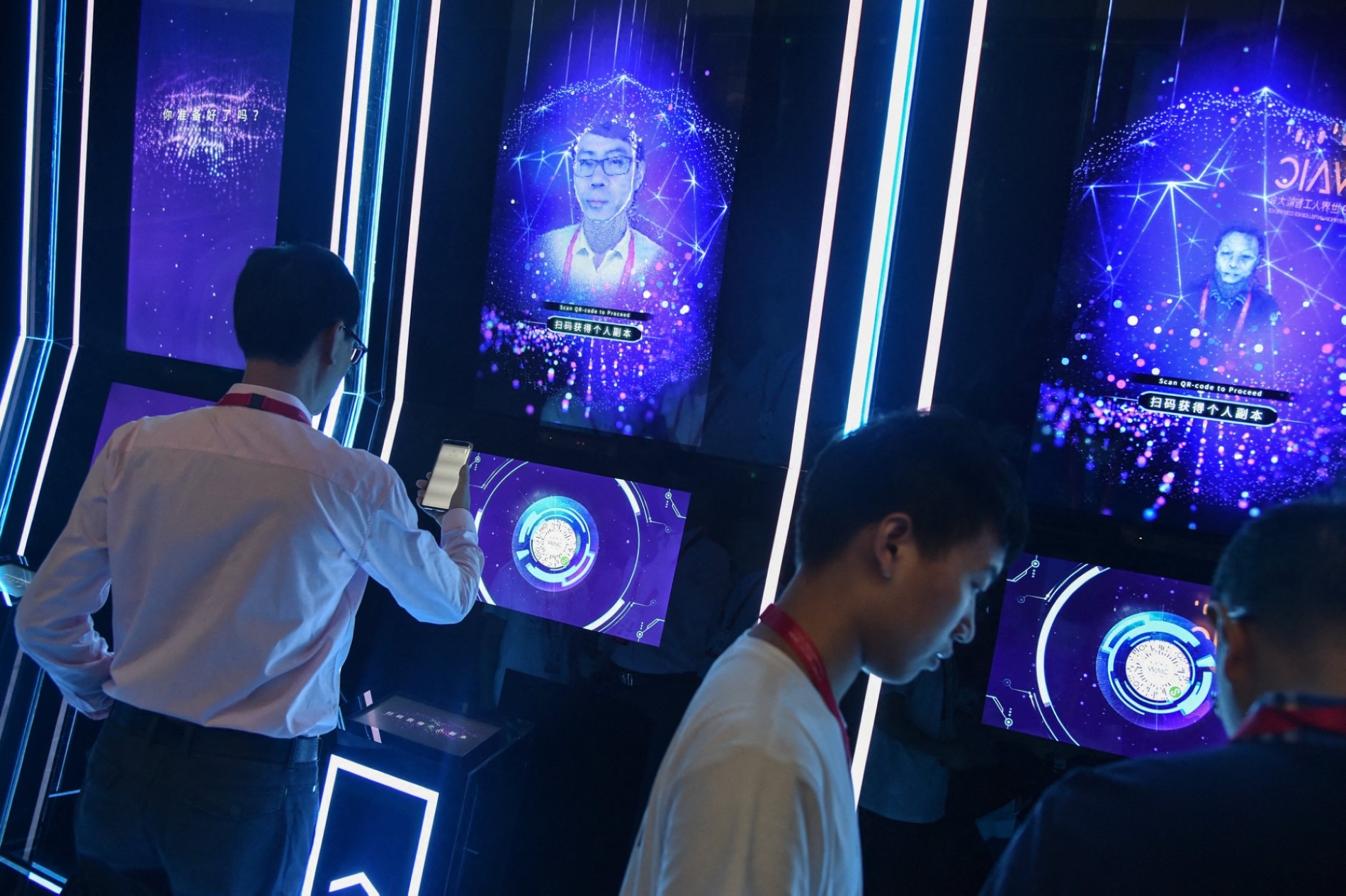
People undergo facial scanning as they arrive for the opening ceremony of the World Artificial Intelligence Conference.
Deep learning
The human eye can recognise changes in facial appearance, such as a drooping eyelid, altered skin colour, or unusual swelling. Other visual patterns associated with health may be too subtle or complex to detect.
Deep learning offers a way to identify these patterns. This branch of artificial intelligence uses multilayered neural networks to extract information from large datasets.
One of the main techniques used in image analysis is the convolutional neural network (CNN). During training, these networks learn to recognise visual features, starting with simple elements such as edges, lines, and colour gradients before progressing to more complex patterns.
Rather than comparing every new photograph directly with all the images encountered during training, the networks learn numerical representations and statistical features. They then use this knowledge to analyse unfamiliar images and generate estimates relevant to the tasks they were designed for.
Vision transformers offer another approach. They divide an image into small patches and examine the relationships between them using a mechanism known as 'self-attention'. This lets the model identify connections between widely separated regions of an image, allowing it to analyse the image as a whole rather than focusing only on individual local features.
One training method involves concealing a substantial proportion of an image, sometimes as much as 75%, and requiring the model to reconstruct the missing portions using the remaining visible information. This technique is one of several possible training strategies and is not essential to every vision transformer.
These developments have expanded the medical applications of image analysis. Researchers are using them to help identify facial characteristics associated with certain genetic syndromes, analyse images of skin conditions and develop digital indicators that may correlate with biological ageing or the progression of particular diseases.
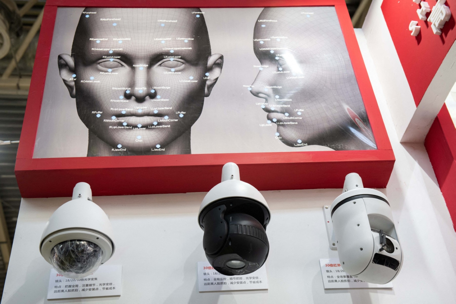
Surveillance cameras featuring artificial intelligence and facial recognition technology are displayed at the 14th China International Exhibition on Public Safety and Security in Beijing.
Biological age
One of the most intriguing applications of facial image analysis is estimating biological age. This reflects the body's condition and physiological functions over time and may differ from chronological age, which simply measures the number of years since birth.
Two people of the same chronological age may have markedly different levels of health, shaped by genetics, environmental influences, lifestyle, and disease. These variations have prompted researchers to develop tools that measure certain aspects of biological ageing.
In a study published in The Lancet Digital Health in 2025, researchers from the US and the Netherlands developed an artificial intelligence system called FaceAge. Designed to analyse facial photographs and estimate age from physical appearance, the system was then used to investigate the relationship between these estimates and survival rates among cancer patients.
To develop the model and conduct initial validation, the researchers used facial images from 58,851 individuals presumed to be healthy, all aged 60 or over. The dataset included 56,304 images for training and an additional 2,547 for initial validation. The team then assessed the model's clinical performance using photographs of 6,196 cancer patients from two medical institutions in the US and the Netherlands.
The findings were also compared with those from a reference group of 535 people without cancer. On average, the model estimated that cancer patients looked 4.79 years older than their chronological age. Patients whose estimated facial age exceeded their actual age by a greater margin tended to have poorer survival rates. The association remained statistically significant even after the researchers accounted for certain clinical and demographic factors.
However, the age estimated by FaceAge is not a direct measure of organ function. Looking older than one's chronological age does not necessarily indicate any particular deterioration in health. Equally, an association between higher estimated facial age and increased mortality risk does not mean that the system can predict when an individual patient will die.
The potential clinical value of such systems lies in the additional information they may offer doctors when assessing a patient's overall condition, alongside physical examinations, laboratory tests and medical history.
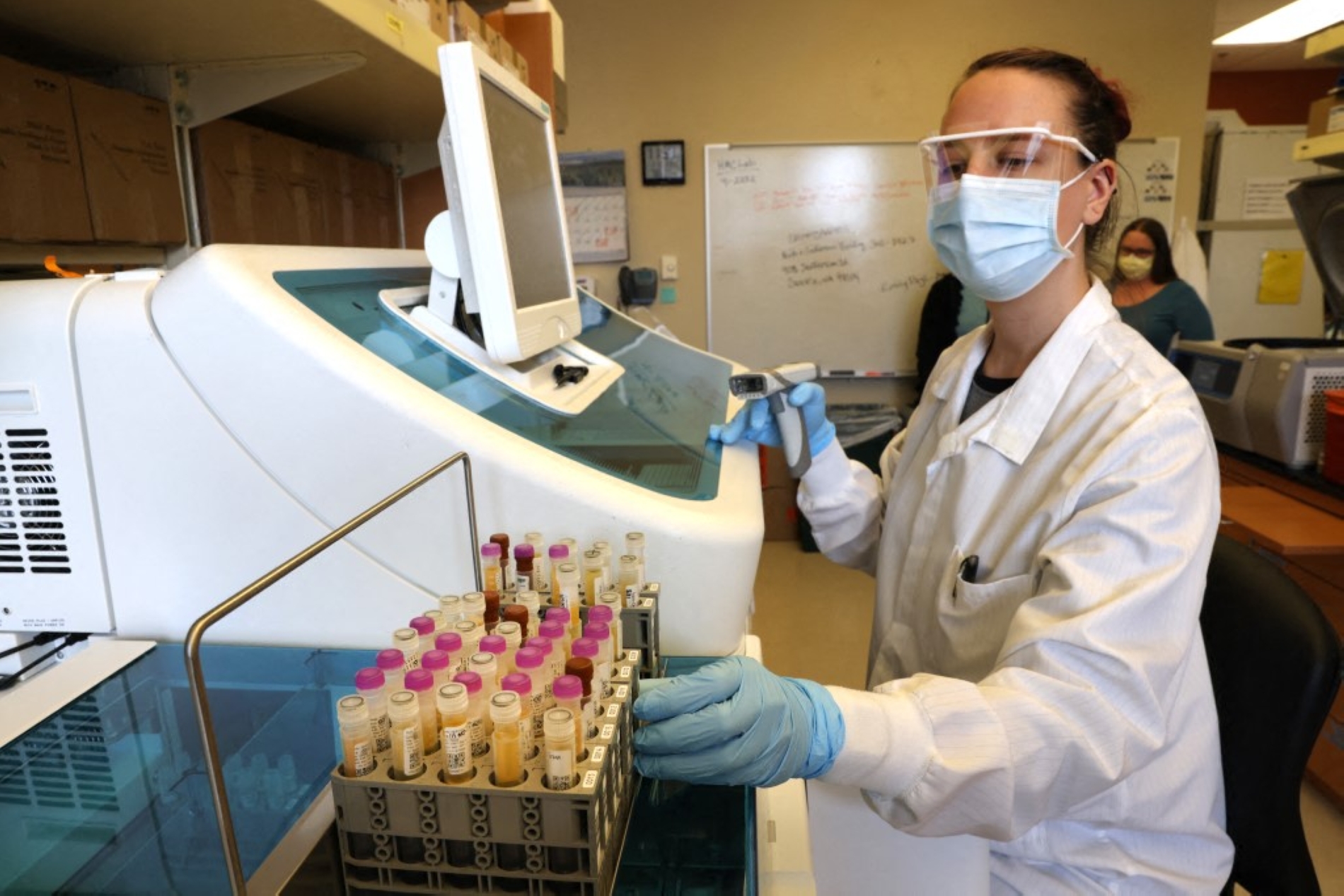
A medical laboratory scientist tests serology samples from the Novavax phase 3 Covid-19 clinical vaccine trial at the UW Medicine Retrovirology Research Lab at Harborview Medical Centre on 12 February 2021, in Seattle, Washington.
Sharpening medical predictions
As artificial intelligence models have become more sophisticated, researchers have turned to larger datasets and developed systems capable of performing several medical tasks using the same facial images.
In a study announced in October, a team from the healthcare and research institution Mass General Brigham, alongside researchers in the US and the Netherlands, developed a system called FAHR-Face. At its heart is a foundation model trained on more than 40 million photographs of human faces.
This approach differs from training a separate model for each task. The system first learns general representations of facial characteristics from an enormous collection of images. It is then adapted to specific purposes using additional data.
The researchers developed two complementary models. The first, FAHR-FaceAge, estimates an indicator of biological ageing from facial appearance, while the second, FAHR-FaceSurvival, predicts mortality risk among cancer patients.
The team used 34,363 photographs of cancer patients to train the second model for its specialised task, then evaluated both models against independent datasets.
The age-estimation model achieved a mean absolute error of 5.1 years when tested on publicly available image datasets, outperforming the reference models included in the comparison. The survival model also distinguished between groups of cancer patients at different levels of mortality risk.
Patients classified in the highest risk quartile had a mortality hazard more than three times that of those in the lowest quartile. After adjusting for clinical factors, the adjusted hazard ratio was 3.23.
This finding does not mean that an individual patient's probability of death is necessarily three times that of another. It describes a statistical association between the model's risk classifications and the rate of deaths during the follow-up period.
The two models also appeared to capture different aspects of risk. Used together, they improved certain measures of mortality prediction compared with either model alone.
Revealing finding
One particularly revealing finding concerned the facial regions the two models relied on when generating their estimates. Although they shared the same underlying foundation model, they focused on different facial features.
Attention maps showed that the age-estimation model placed greater emphasis on the nasolabial folds, the creases running from either side of the nose towards the corners of the mouth, as well as the forehead and temples. The mortality risk model assigned greater weight to the areas beneath the eyes and around the bridge of the nose.
This difference suggests that the visual patterns associated with ageing may diverge, at least in part, from those linked to mortality risk among cancer patients.
Identifying the regions that influence a model's predictions does not, however, explain the biological processes underlying these associations. Nor does it establish that visible changes in those areas cause an increased risk of death.
The researchers' tests suggested that both models maintained their performance across groups differing in age, sex, race, and cancer type. They also demonstrated resilience to certain common variations in photographs, including changes in lighting, camera angle, and the use of cosmetics.
Successful performance on evaluation datasets nevertheless does not establish that the models will work equally well across all populations or in every clinical setting.
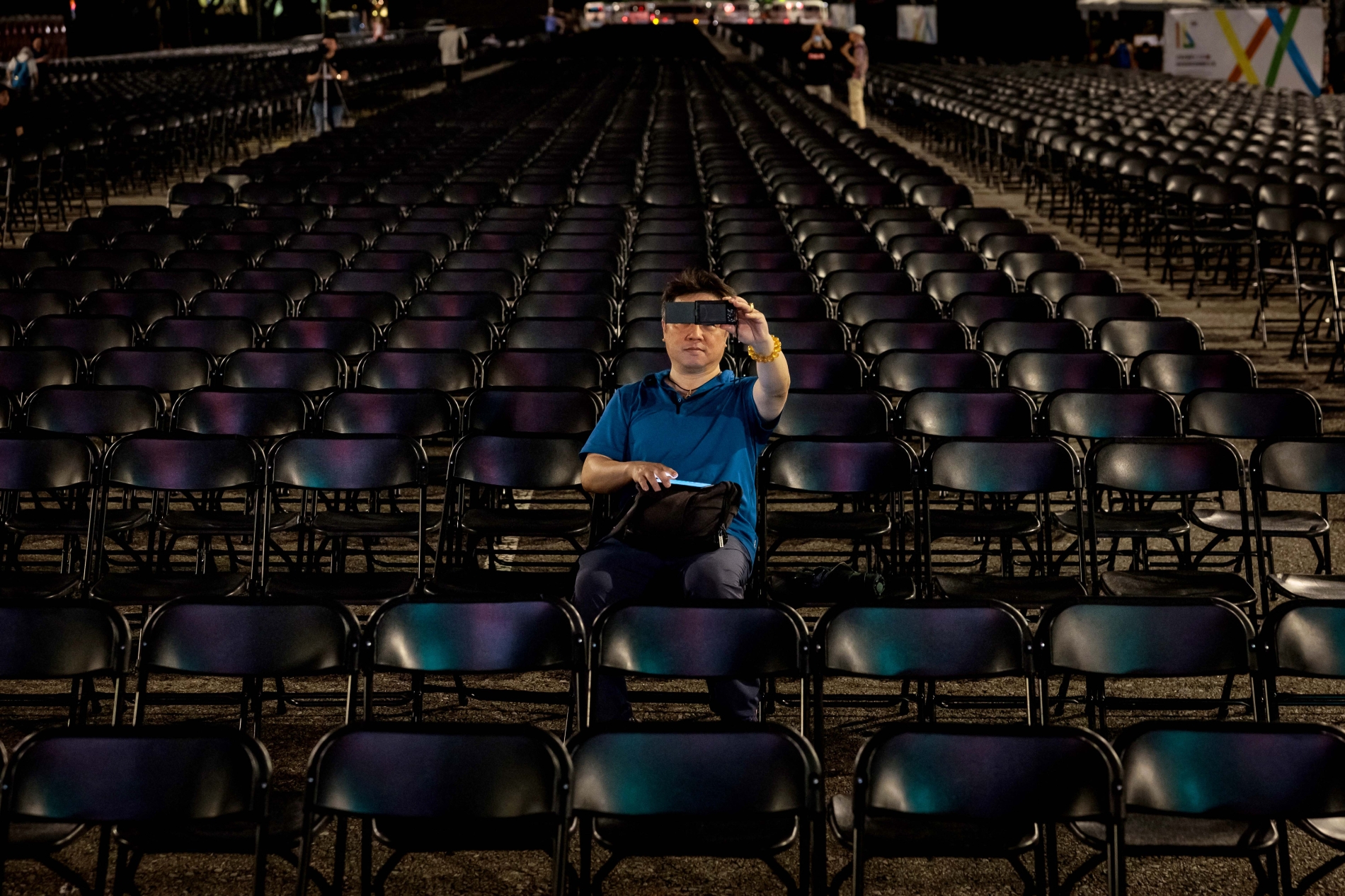
A man takes selfies with chairs installed for the inauguration ceremony of Taiwan's President-elect Lai Ching-te and Vice President-elect Hsiao Bi-khim in front of the Presidential Office in Taipei on May 19, 2024.
Substantial challenges
Despite their promise, these technologies face substantial scientific and ethical challenges before they can become part of routine hospital practice.
Facial photographs are influenced by numerous factors, including lighting, camera quality, facial expressions, and the natural changes that accompany ageing, as well as differences between individuals and populations.
Using personal photographs for medical analysis also raises important questions about privacy and data protection, particularly because facial images constitute biometric information that can identify individuals.
Researchers must establish whether the information extracted from photographs offers genuine benefits beyond existing clinical assessment tools. Crucially, they need to show that using these systems improves medical decisions or patient outcomes, rather than merely identify statistical associations through retrospective analysis.
Accurate predictions of mortality risk in research datasets do not guarantee of better treatment or longer lives once a model enters clinical practice
Accurate predictions of mortality risk in research datasets do not guarantee of better treatment or longer lives once a model enters clinical practice.
The technology may prove most useful in helping doctors assess patients' overall condition, particularly where certain examinations are difficult to obtain or medical resources are limited. Clinical expertise and established diagnostic procedures would remain essential.
For Troutwine, a selfie revealed an unusual symptom that prompted her to seek medical advice. In research laboratories today, scientists are pursuing a more ambitious goal: turning facial features into digital indicators that could offer a deeper understanding of human health.
However, an essential distinction between recognising a symptom in a photograph and using an algorithm to assess medical risk. Facial images may provide valuable clues, and artificial intelligence may eventually help doctors interpret them. Yet a photograph cannot become a reliable diagnostic instrument solely because an algorithm has learnt to extract patterns from it.